Lung sliding
By erich | Last updated: Apr 29, 2026
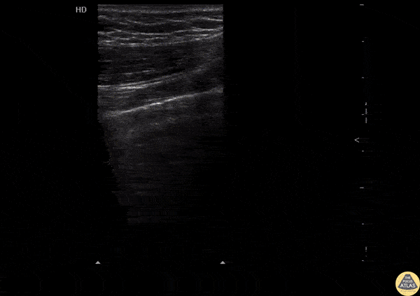
Visceral and parietal pleura in direct contact and moving against one another causes a shimmering "ants crawling on a line" appearance.
This is best seen with the depth and gain down with multi beam and smoothing disabled.
Linear high frequency probes are best, but any probe will do. Have the probe oriented longitudinally (cephalo-caudad).
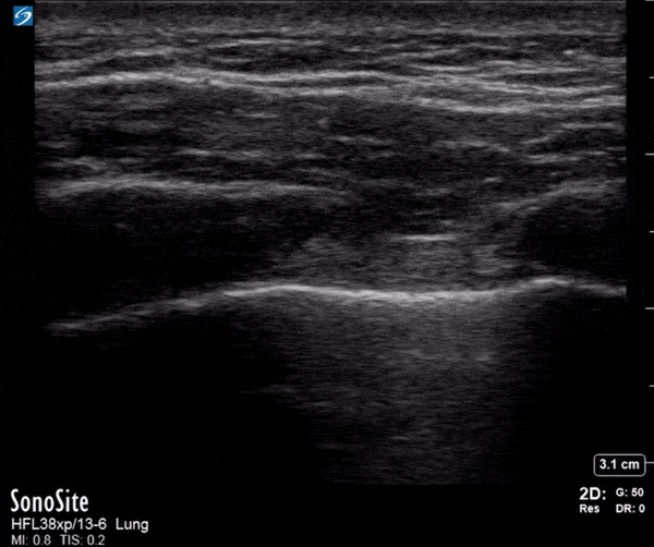
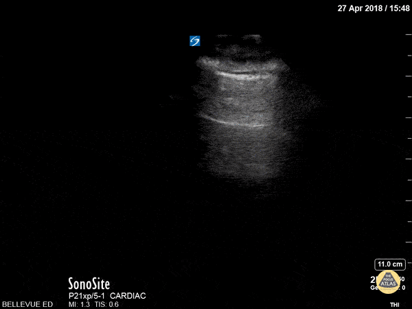
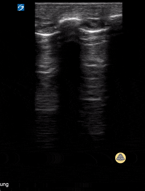
If unsure of the visual motion M-mode is useful:
Seashore Sign confirms normal lung sliding. The static chest wall looks like straight lines (the sea), and the moving lung looks like sand (the beach). Barcode (Stratosphere) Sign shows absent lung sliding due to apnoea, misplaced endotracheal tube, pneumothorax, pleural or lung pathology. The entire image consists of horizontal lines because there is no motion beneath the parietal pleura.
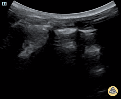
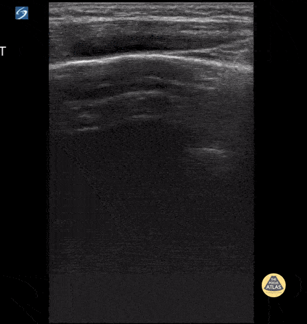
Normal Lung sliding
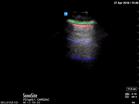
Lung sliding - Colorised green: subcutaneous tissue, red: pleural space, blue: a lines
Illustrating improved lung slide visualization by decreasing the gain
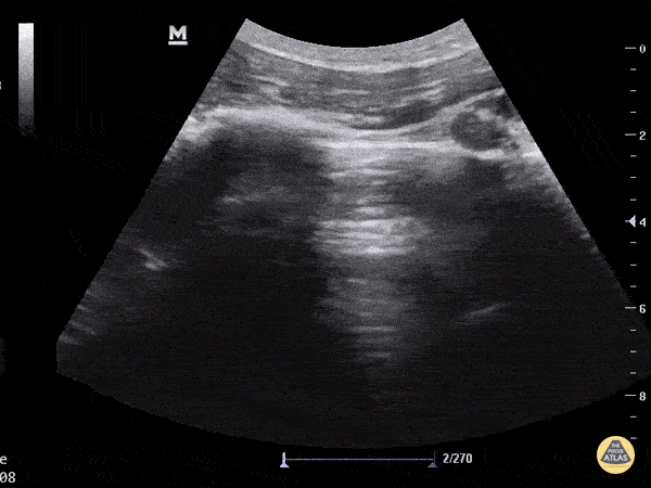
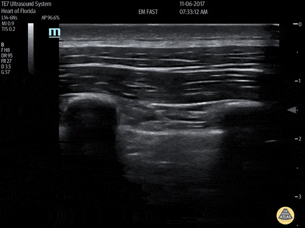
Normal lung sliding - FAST in trauma case
No lung sliding
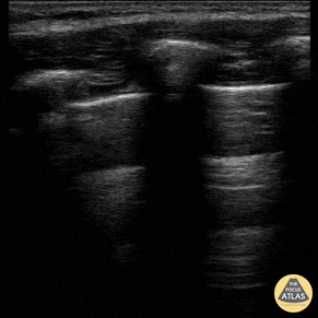
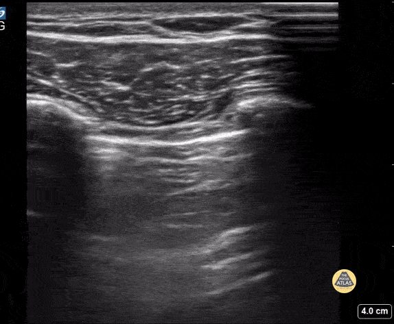
Lung sliding and A-lines
Normal Lung sliding in paediatric (21 month) trauma case
Normal Lung sliding during FAST exam in 11 year old
Normal lung sliding with comet tails in 4 year old
No lung sliding in paediatric case
Lack of lung sliding in paediatric case
Reference
Comments
Make a CommentNo comments yet. Be the first to comment!